
Consultant Breast & General Surgeon
BMI Chiltern, BMI Shelburne &
Buckinghamshire Hospitals NHS Trust
BREAST SURGERY
ONE-STOP CLINICS
BREAST CANCER
RECONSTRUCTION
BREAST AUGMENTATION
DAY SURGERY
HERNIA REPAIRS
GENERAL SURGERY
© Mr Giles Cunnick 2008 / All rights reserved
Web Site Designed by www.appliedwebsites.co.uk
Breast Clinics
Breast Clinic Information
The aim of the breast clinic is to provide the highest quality diagnostic service to patients with breast complaints, ranging from simple benign problems to breast cancer. Urgent referrals from GPs are always seen within 2 weeks, and often much sooner. Each patient is assessed and treated individually with a full complement of specialised staff, modern equipment and state-of-the-art services. The following summarises the various aspects of care offered at the clinic.
Out-Patient Clinic Pathway
Patients with breast lumps, pain, discharge and other breast symptoms are seen in a ‘one-stop clinic’. Initially, patients are either seen by myself or undergo breast imaging beforehand (mammograms and/or ultrasound scan). Once all the scans have been reported by the Consultant Radiologist, the patient will be told the results on the same day. If any abnormality is detected, it may be necessary to perform further investigations, such as further mammograms or a needle test/biopsy. These tests are also usually performed on the same day. In this way, a diagnosis can be established in a single visit for most women attending the clinic. A specialist Breast Care Nurse is also present to give further information and support to the patient, if desired. She will also chaperone the patient during the examination.
The principal diagnostic tests are outlined below:
Mammography
Mammography is a way of imaging the breast tissue and produces an X-ray called a mammogram.
Each breast is compressed between two plastic plates while a beam of X-rays passes through the breast tissue onto an X-ray film.
Two different views of each breast are taken. The process may be slightly uncomfortable but only lasts 30 seconds.
The mammograms are then immediately reported by the Consultant Radiologist who has specialist expertise in breast radiology.
Ultrasound
Ultrasound machines use sound waves to image various areas of the body, including the breasts. The sound waves are transmitted through a probe, which is placed directly over the breast. Lubricating gel is placed on the breast skin to improve contact. The sound waves are reflected within the breast back to the ultrasound machine, which transforms them into a computer image viewed on a display monitor.
The ultrasound examination is carried out by the Consultant Breast Radiologist, who decides whether any abnormalities are present.
The test is very good at differentiating between solid lumps and fluid-filled lumps (cysts). It is particularly useful in women under the age of 35, since younger breasts are much denser; this means that abnormalities may be sometimes difficult to visualise on mammograms in this age-group. Ultrasound examination does not usually cause any pain
FINE NEEDLE ASPIRATION CYTOLOGY |
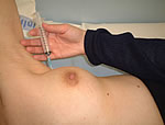 |
The cells are smeared onto a microscope slide and sent to the laboratory to be analysed by an experienced Consultant Pathologist who specialises in breast pathology.
This test is highly sensitive at distinguishing between benign and malignant cells. Furthermore, it can be used to aspirate fluid from a cyst, following which the cyst disappears. It is slightly uncomfortable and sometimes causes bruising.
This test is performed under a local anaesthetic using a wider needle to allow a larger number of cells to be examined by the Consultant Pathologist. It may cause more bruising than the fine needle test. It is usually performed under radiological guidance (i.e. during an ultrasound or mammogram). Although the test often gives a definitive diagnosis, it takes a few days before a result is available due to laboratory processing. Appearance of a breast core biopsy under the microscope.